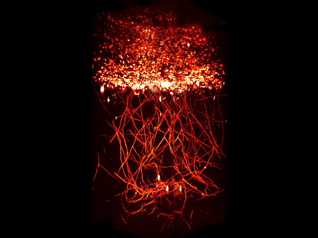
Deep Vascular Imaging in Wounds
Here you’ll see a really nice example of what modern microscopy actually allows you to do if combined with additional, fitting, techniques
When we’re wounded, our bodies rush to repair the damage. First, there’s a blood clot that forms quickly and acts as a temporary seal. Then follows the more gradual process of angiogenesis – the growth of new blood vessels to replace those that are injured. This image is of a wound healing on a mouse. It was captured using a new form of fluorescence microscopy, which involves staining the tissue with chemicals that make it fluoresce, and then taking a picture that shows only the fluorescence. At the top is the blood clot in the open wound, while the strands beneath are newly grown capillaries [the smallest blood vessels] extending into the muscle. This technique enables us to understand the complex mechanisms of wound healing better than ever before.
Abstract
Deep imaging within tissue (over 300 μm) at micrometer resolution has become possible with the advent of…
View original post 4,371 more words


Leave a comment